- Home
- Health Science
- Feeding the gut microbiome: what fibre research can and cannot promise
Feeding the gut microbiome: what fibre research can and cannot promise
Food, microbes and evidence
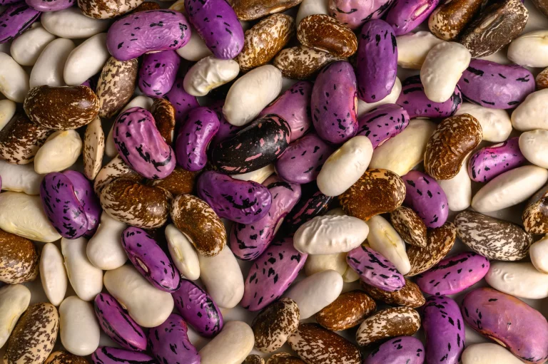
A serving of beans is familiar food. The activity it supports inside the gut is much less visible. Researchers are studying how food components interact with communities of microorganisms, and how those interactions influence the substances our bodies encounter. That research is promising, but it does not turn every claim about gut health into established medicine.
The useful question is more specific than whether a food is good for the microbiome. Which component changed, in whom, over what period, and with what consequence? Looking at those details helps separate a biological finding from a promise on a supplement label.
A controlled study offers a closer look
In a 2023 research update, the US National Institute of Diabetes and Digestive and Kidney Diseases described a randomized feeding study involving 17 healthy, weight-stable adults. Participants received a diet designed to nourish gut microbes and a more typical Western diet, each for approximately three weeks.
The researchers used a metabolic ward to examine energy intake, expenditure and output, comparing each participant across the two diets. They reported changes in microbial composition and function, a small reduction in body fat stores, and differences in hormones associated with fullness.
Those findings are interesting partly because the conditions were carefully measured. They are also limited: a small, short study in healthy adults cannot establish long-term weight loss, show that every person will respond similarly, or prove that a marketed product recreates the dietary intervention. The research is a starting point for better questions, not a complete household prescription.
Another distinction matters: the diets differed in several ways. A comparison between dietary patterns cannot by itself assign every observed change to fibre alone, or establish which particular food was responsible.
Microbes can change what food makes available
A separate NIDDK update from 2022 described work on orange fibre and a molecule called N-methylserotonin. Experiments in mice identified a role for particular gut bacteria in releasing the molecule from the fibre. A small study in women also found evidence that this release could occur in humans.
The animal experiments explored effects on body fat and metabolism, but those outcomes should not be carried across to people as proven benefits. Demonstrating that a compound appears in human stool is different from demonstrating that it treats a disease, produces sustained weight loss or is safe and effective as a supplement.
Graymedy's interpretation is that these studies illustrate a relationship: food structure, microbial activity and human biology interact. They do not identify one ideal microbial community for everybody. Nor do they justify describing an ordinary meal as a treatment that has been clinically tested in all readers.
Food and probiotic products answer different questions
Dietary fibre is not the same thing as a probiotic. The National Center for Complementary and Integrative Health describes probiotics as live microorganisms intended to provide health benefits. Products can contain different organisms, and an effect demonstrated for one type does not automatically apply to another.
NCCIH also notes important gaps in evidence and safety knowledge. People with serious illnesses or weakened immune systems can face greater risks, and probiotic products should not replace proven treatment or delay assessment of a health problem. A supplement decision therefore deserves its own conversation with a healthcare professional.
When reading a gut-health claim, ask whether the evidence concerns a whole diet, an isolated fibre, a bacterial strain or a finished commercial product. These are distinct interventions. A product that borrows the vocabulary of a study still needs evidence that matches what it actually contains and promises.
Keep everyday choices proportionate to the evidence
NIDDK's patient information identifies whole grains, legumes, fruits, vegetables and nuts as fibre sources. It advises increasing fibre gradually and discussing an appropriate intake with a health professional. Fluid needs should also reflect a person's circumstances and health, rather than a single rule copied from a headline.
For a practical discussion, start with foods already available and affordable: could a meal include lentils, beans, oats or vegetables that fit the person's dietary needs? This is an editorial prompt for planning, not an instruction to override restrictions prescribed for a medical condition.
The most credible promise of microbiome science is better understanding of why responses differ. Readers can value that progress while remaining cautious about claims of a quick reset, a universal bacterial score or guaranteed results. A useful article should leave the uncertainty visible alongside the discovery.
Sources: NIDDK: Controlled feeding study (2023); NIDDK: Orange fibre research (2022); NCCIH: Probiotics usefulness and safety; NIDDK: Fibre and nutrition.
Editorial review: 20 October 2026. Educational explainer based on the cited sources; no original interviews or individual medical advice.
Image: Jana Ohajdova / Pexels, used under the Pexels License. Illustrative stock photograph; no endorsement or individual diagnosis is implied.


Health Science
View more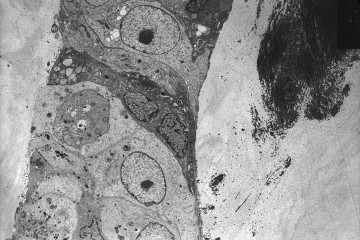
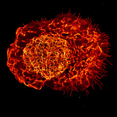
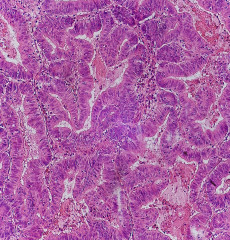
A new environmental-health finding is more specific than a broad claim that a chemical causes breast cancer. NIH researchers reported an association between two legacy PFAS and estrogen receptor-negative breast cancer, while finding no clear association with breast cancer overall. The finding behind the headline NIH’s November 5, 2026 report describes research from the Sister Study. Investigators measured 17 PFAS in blood collected before diagnosis, comparing 1,609 women who developed breast cancer with a comparison sample of 2,269 women. Participants had a sister with a previous breast-cancer diagnosis. Women in the highest PFOA group had a 67% higher rate of ER-negative breast cancer than those in the lowest group. For PFOS, the relative difference was 93%. These are comparisons between exposure groups, not an individual woman’s absolute probability of developing cancer. What it does not show An observational association does not, by itself, establish causation. The analysis also does not show that every PFAS has the same relationship with cancer. The authors called for research on newer replacement compounds and on populations with higher exposures. For readers, three questions help keep the result in proportion: which exposure was measured, which outcome was associated with it, and which people were studied? Replacing those details with a general claim can make an informative finding misleading. Keeping uncertainty useful Graymedy’s interpretation is that a specific signal is a reason for further investigation, not a reason to assign blame for an individual diagnosis. Environmental-health evidence is most useful when it helps define the next study and supports clear public communication. It should also be possible to discuss uncertainty without dismissing the concern. Replication, better exposure measurement and examination of potential mechanisms can strengthen or revise the picture. The appropriate response to this paper is careful attention rather than a promise of a new screening test or treatment. Sources NIH research report, November 5, 2026; Chang et al., Journal of Clinical Oncology. Editorial review: November 9, 2026. Independent research analysis; not individualized risk assessment. Image: National Cancer Institute, ID 2175, archival breast-cancer cell micrograph, 1985. Public domain. Image source. Not a Sister Study specimen or PFAS experiment.
Depression research finds a possible bottleneck in the brain’s memory centre
Can changing scarred tissue help cancer immunotherapy work better?
Christopher Rodell is testing whether a gel can teach the immune system to accept new beta cells
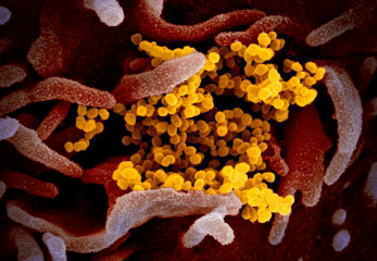
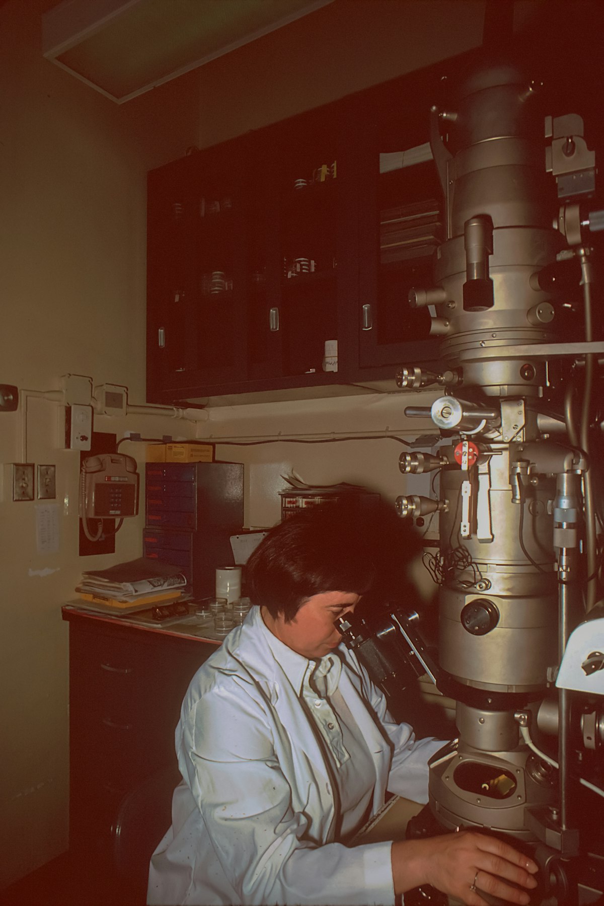
A virus’s genetic material is often pictured as a set of instructions waiting to be copied. New coronavirus research suggests a more active role: particular stretches of RNA help organise contacts with viral proteins, and disrupting those contacts can interfere with replication. That makes the meeting points themselves interesting places to look for future medicines. A study highlighted by UC Riverside on 30 October and UC San Diego on 2 November 2026 maps these interactions across SARS-CoV-2. The experiments identify biological vulnerabilities in cultured cells. They do not announce an antiviral ready for patients or establish protection against a future pandemic. Reading the genome as a working structure RNA can carry genetic information, but it also has structural and regulatory functions. The National Human Genome Research Institute describes several forms of RNA with different cellular jobs and notes that some viruses use it as their genome. A sequence can therefore be both information and part of an operating mechanism. This study examines that second possibility in the coronavirus life cycle. Instead of looking only at a protein as an isolated target, the researchers asked where proteins contact the viral RNA. The resulting map offers a way to study how those contacts influence the virus’s activity. The shift is useful because a genome’s sequence alone does not explain everything it does. Knowing the letters is different from understanding which molecular partners use them, when those partners bind and what happens if access is blocked. Finding contacts, then testing their importance The team used enhanced cross-linking and immunoprecipitation, usually called eCLIP, to survey interactions involving three viral proteins: NSP8, NSP12 and nucleocapsid, or N. The approach looked across the genome rather than limiting the search to sites already expected to be important. UC San Diego’s account describes an interaction between N and a region near the genome’s beginning, the 5′ untranslated region, with a role in protein production. It also describes a previously unrecognised interaction between NSP12 and RNA encoding another viral protein, NSP3. These findings give researchers more specific functions to investigate. A binding map is a starting point. The stronger question is whether interfering with a contact changes what the virus can do. This is where the study moves from description towards a functional test. Blocking a meeting point The investigators used short, modified nucleic-acid sequences designed to bind selected RNA regions and obstruct their interactions. Blocking the sites reduced viral replication in Vero cells, which originate from African green monkey kidney cells. Similar effects were observed in human lung cells. Those are cell-culture experiments. UC Riverside explicitly reports that animal work is still needed to determine whether the findings could translate into a therapeutic approach. Human cells in a laboratory are biologically informative, but they do not reproduce an entire person’s infection, immune response or drug exposure. The distinction matters for the next stage of research. A candidate would need to reach the relevant cells, remain active there and avoid unacceptable effects. Identifying a vulnerable interaction and delivering an effective treatment against it are connected but separate achievements. Could the approach outlast viral change? The researchers are interested in contacts involving machinery or sequences shared among coronaviruses. If an interaction is essential and difficult for a virus to alter, it could be a more durable target. That is the rationale for broader antiviral development, rather than evidence that resistance has already been overcome. Graymedy’s interpretation is that breadth should be tested directly. A promising result against one virus cannot establish the same effect against a related virus, and similar sequences do not guarantee identical biological behaviour. Studies across viruses and experimental settings would help define the actual reach of the idea. The same care applies to comparisons with existing medicines. Current antivirals already target essential viral processes. This work adds possible points of intervention; it does not show that a new approach is safer or more effective than treatments used today. Why this map is worth following The contribution is a more detailed view of how viral components cooperate. It produces testable questions about regulation, replication and vulnerability, giving later drug-development work something more precise than a broad instruction to attack the virus. Its importance will grow if those questions lead to reproducible effects in more realistic models and eventually useful clinical candidates. For now, the breakthrough being pursued is an understanding of viral machinery that could widen future treatment options, with the distance to patient benefit kept visible. Sources: UC San Diego: research report, 2 November 2026; UC Riverside: methods and limitations, 30 October 2026; Study record: genome-scale eCLIP mapping, Nature Communications; NHGRI: RNA and its functions. Micrograph: NIAID-RML / Wikimedia Commons, CC BY 2.0; resized. Reviewed 4 November 2026. Independently written from public sources; no interview was conducted. General information, not individual medical advice.
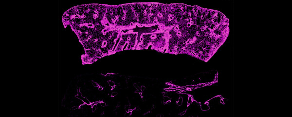
Transplant research looks at the drainage system around the immune response
A new colorectal cancer atlas looks beyond the under-50 divide
Why Replication Matters More Than a Single Breakthrough Study
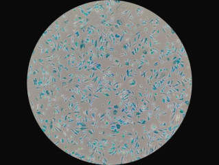
Some cells stop dividing without disappearing. They remain biologically active, sending signals into surrounding tissue. This state, called cellular senescence, has become a major focus of ageing research because it can help protect the body in some circumstances and contribute to damage in others. The challenge is telling those situations apart. Researchers need to know which cells have become senescent, where they sit, and what they are doing before they can design treatments that target them accurately. A growing effort to map these cells is making that question more concrete. Why a stopped cell can still matter Senescence is a response to stresses such as DNA damage or disrupted cell function. Stopping a damaged cell from multiplying can provide an important safeguard against cancer. Senescent cells also participate in normal processes, including tissue repair. Their presence alone therefore does not automatically mean disease. Problems can emerge when these cells persist and release substances that alter their surroundings. Scientists call this mixture the senescence-associated secretory phenotype, or SASP. Its components can influence inflammation, neighbouring cells and tissue maintenance. The effects depend on the biological setting, which makes the familiar description of senescent cells as simply harmful incomplete. A SenNet Consortium perspective in Nature Aging explains why cell identity, the trigger for senescence and the tissue environment all matter. A single measurement cannot capture that complexity across the whole body. A map built around cellular differences In June 2026, the US National Institutes of Health reported a collection of studies advancing the Cellular Senescence Network, known as SenNet. The research introduces a framework of “senotypes”: patterns that distinguish senescent cells according to their context and characteristics. The work includes mapping in tissues such as the lungs, lymph nodes and the brain’s prefrontal cortex, alongside methods for detecting rare cells within complicated tissue samples. Combining molecular measurements with information about location helps researchers investigate how cells interact with their immediate neighbourhood. As the NIH announcement makes clear, this is groundwork for more selective therapies. An atlas can help researchers identify promising targets; it does not itself establish that a treatment prevents age-related illness. What blood measurements may reveal A related NIH research summary describes a tool called the Senescence Catalog, or SenCat, built from gene activity and protein measurements in 14 types of human senescent cells. The findings reinforce that senescence has several molecular forms rather than one universal signature. In companion research, investigators measured associated blood proteins in more than 2,000 people from two long-term ageing studies. Some patterns were linked to health characteristics including kidney disease, walking pace and diabetes. Cell-specific signals also appeared relevant: markers connected with kidney-cell senescence were associated with kidney disease. These reported associations make the proteins useful research leads. They do not show that a commercial blood test can reliably guide an individual’s treatment, or that changing a marker will necessarily improve health. Why removing every senescent cell is not the goal Experimental medicines called senolytics aim to eliminate selected senescent cells. Animal research has encouraged interest in this approach, but translating it into useful care requires a careful balance. The same broad cellular process can contribute to disease while also supporting healing and limiting uncontrolled growth. The NIH Intramural Research Program describes efforts to find molecular markers that distinguish relevant cells and measure whether interventions affect them. Identifying a target, reaching it safely and demonstrating meaningful benefit are separate tasks. Results in mice cannot establish that an intervention will extend healthy life in people. Future clinical studies will need to establish who benefits, which conditions should be treated and how outcomes change over time. Tracking side effects will be as necessary as measuring changes in cell numbers or blood proteins. What progress should look like SenNet’s public programme is designed to share maps, methods and common definitions so that laboratories can compare results. That infrastructure matters because inconsistent ways of identifying senescent cells can make apparently similar studies difficult to interpret. For readers, the useful questions are specific: which cells were studied, in what tissue, and was the evidence obtained from laboratory models or people? A credible treatment claim must also show a measurable health benefit and an acceptable safety profile. The promise of this research lies in understanding cellular ageing precisely enough to intervene wisely, while preserving the functions that help keep tissues healthy.

Sleep and the Brain’s Waste-Clearance System: What Human Studies Reveal

Your Body Has Multiple Clocks: How Circadian Rhythms Shape Sleep and Metabolic Health