- Home
- Health Science
- Human lung organoids reveal a local immune response to viruses
Human lung organoids reveal a local immune response to viruses
Lung immunity research
Small pieces of human lung tissue grown in the laboratory may offer researchers a new way to study how the lungs respond to viruses. An NIH-supported team has developed organoids that retain immune cells alongside other tissue components, allowing the researchers to observe local antiviral responses.
In the study, reported in October 2026, researchers used lung tissue from more than 220 volunteers and maintained the models in a three-dimensional air–liquid culture system for up to several months. The organoids retained several immune cell populations, including tissue-resident T cells.
Testing a response
When the researchers exposed the models to SARS-CoV-2, they observed virus-specific adaptive immune responses. Pre-stimulating the organoids with viral fragments reduced subsequent infection in the laboratory model. The findings support further investigation of local lung immunity and of vaccination strategies aimed at mucosal defenses.
The work is a research advance, not a new vaccine or a clinical treatment. An organoid cannot reproduce the full complexity of a person’s body, and results in tissue culture do not establish how a vaccine will perform in people. Further studies are needed to translate the findings.
Why the model matters
Because the culture retains tissue and immune components together, it gives scientists a way to investigate interactions that simpler cell models may miss. The researchers suggest it could also help study other respiratory diseases in the future, although those uses remain to be established.
What makes this model different?
Many laboratory studies use isolated cells to answer a precise question. That approach is useful, but it can remove cells from the surroundings in which they normally interact. The Stanford team’s model used intact fragments of distal human lung tissue in a three-dimensional air–liquid interface. NIH says the culture system simulated aspects of the lung’s gas-exchange environment and maintained the tissue for months.
The organoids retained structural components and several immune populations. In addition to T cells, the published abstract reports B cells, natural killer cells and myeloid cells. Retaining these populations creates an opportunity to observe responses within a tissue setting rather than examining just one type of cell in isolation.
One focus was tissue-resident memory T cells. These immune cells remain in tissue and can respond when they encounter a recognized pathogen again. The researchers examined whether such cells in their lung models retained the ability to recognize viruses and whether a local response could limit infection.
From a viral challenge to a research question
In the SARS-CoV-2 experiments, the team observed responses specific to the virus, alongside more general inflammatory reactions. In a separate step, the organoids were exposed to viral fragments before infection. The later reduction in infection provided evidence that local immune activity in the model could contribute to protection.
This is a controlled laboratory experiment, not a trial in volunteers given a new vaccine. The tissue fragments came from volunteers, but the experimental exposure occurred in culture. The study cannot establish how an intervention would perform in a whole person, how long protection would last, or whether it would be safe and effective in a clinical population.
Where the work could lead
NIH says the platform may help investigators study local tissue immunity separately from other parts of the immune system. That separation is an advantage for some questions and a limitation for others: the circulation, other organs and the full immune system are not reproduced in a dish. Researchers will need to compare observations from the model with other experimental systems and, eventually, appropriate clinical evidence.
The authors suggest future applications in respiratory conditions such as asthma, chronic obstructive pulmonary disease and bacterial infection. Those are proposed directions, not findings that this study has already established for each disease. Different pathogens and conditions may require different experiments and validation.
The immediate contribution is a research platform and evidence about lung-resident immune responses. Whether it can guide a new local vaccination strategy will depend on additional studies of the mechanism, reproducibility and real-world clinical outcomes. The distinction between a promising model and a proven intervention is essential when interpreting this work.
Sources: NIH research release; Rathkey and colleagues, Nature, 2026.
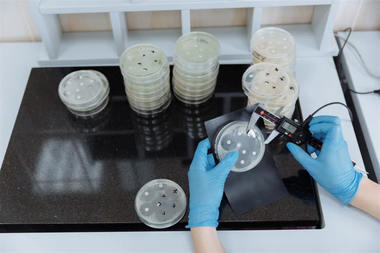
Featured image: illustrative laboratory culture measurement by Thirdman / Pexels; it does not show the organoids in the study.

Health Science
View more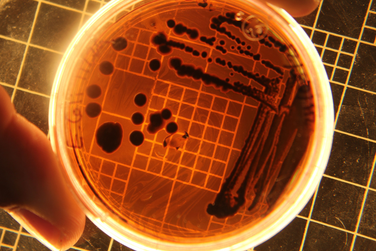
Antibiotics can be lifesaving when a bacterial infection needs treatment. Their effects, however, are not confined to the harmful bacteria causing an illness. The medicines can also change the communities of microorganisms that normally live in the gut. Scientists call this community the gut microbiome. It includes many kinds of bacteria and other organisms, and it varies from person to person. Researchers are studying how it changes after antibiotics, how it recovers and why some changes last longer than others. Following recovery over time In a 2022 study published in Cell Reports, researchers followed healthy adult volunteers before, during and for six months after exposure to four commonly used antibiotic regimens. They analysed stool samples to track bacterial species, genes associated with antibiotic resistance and other features of the microbial community. The researchers observed an immediate fall in the number of bacterial species and in bacteria that could be cultured. For most participants, species richness—a count of different species—returned to its pre-treatment level after two months. That did not mean the community was identical to the one present before treatment. Its composition, resistance-related genes and metabolic output could still differ. Some volunteers had a more persistent reduction in diversity. The study also found differences between antibiotic regimens, including slower recovery of species richness after azithromycin. These findings describe patterns in the study group; they cannot predict exactly what will happen to a particular person after a prescription. What is the resistome? The term “resistome” refers to the collection of antibiotic-resistance genes found in a microbial community. Antibiotics can favour bacteria that survive their effects. Resistance is a property of microbes, not a change that makes the human body itself resistant to a medicine. Resistance genes can be found in bacteria that are not currently causing illness as well as in pathogens. Their presence is important to researchers, but a laboratory finding in a stool sample does not automatically mean a person has a resistant infection or needs treatment. What the study cannot tell us The volunteer study was small and involved healthy adults rather than people being treated for serious infections. Different ages, illnesses, diets, medicines and previous antibiotic exposures can affect the microbiome. A later study using prescription records and stool data from nearly 15,000 Swedish adults examined associations between prior antibiotic use and microbiome composition, but observational associations alone cannot establish every cause. Scientists still need to understand which changes have meaningful health consequences, who is most vulnerable and which interventions are helpful. No single measure, including species richness, captures the full function of this complex ecosystem. Claims that a product will reliably “reset” everyone’s microbiome go beyond what these studies establish. Practical care still matters The findings support careful antibiotic use, not avoidance of necessary treatment. Antibiotics do not treat viruses that cause colds and many other respiratory illnesses. But when a clinician determines that an antibiotic is needed, the benefits usually outweigh the risks. Take it as prescribed and ask the prescriber or pharmacist about side effects and any concerns. Do not save leftover antibiotics, share them or use them for a later illness. If symptoms worsen or a troublesome reaction occurs, contact a health professional. Severe symptoms need urgent care. Ask before taking a probiotic or supplement, especially if you have a serious illness or weakened immune system; the evidence and suitability differ by product and person. The gut microbiome can be resilient, but recovery is not always a simple return to the starting point. That is one reason using antibiotics thoughtfully matters for both individual care and the wider problem of antimicrobial resistance. Sources: Anthony et al., Cell Reports, 2022; Swedish population study; CDC antibiotic guidance. Photo: Wassily Kandark / Pexels. Illustrative bacterial culture, not a sample from the studies. https://www.pexels.com/photo/bacterial-colonies-shown-under-a-light-19728524/
Human lung organoids reveal a local immune response to viruses
A1C and blood glucose: why diabetes tests tell different stories

Why Can You Sleep All Night and Still Wake Up Tired?
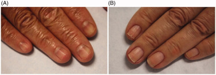
A nail that splits, peels or breaks easily can make everyday tasks frustrating. It is also easy to find confident claims online that a particular nail appearance reveals a particular deficiency. In practice, the picture is rarely that simple. A surface with a history Nails are made mainly of keratin. The visible nail plate grows gradually from the nail matrix near its base, so changes can remain visible as the nail grows out. Repeated wetting and drying, contact with irritating substances and physical wear may all contribute to fragile nails. A clinician considers the pattern as well as the appearance: whether one nail or several are affected, how long the change has been present, and whether there has been injury, skin disease, medication use or other health changes. A nail alone cannot reliably diagnose a nutrient deficiency. Care for everyday wear Gentle nail care, moisturising hands and nails, and protecting hands when they are repeatedly exposed to water or cleaning products may help reduce everyday stress. Avoid picking or aggressively cutting the skin around nails. If you use nail products, consider whether a change in your routine coincided with irritation or damage. Supplements are not automatically the answer. Discuss persistent concerns with a healthcare professional, who can decide whether tests or treatment are appropriate. When to have a nail checked Seek medical advice for a new or changing dark streak, a nail separating from the skin, pain, swelling, bleeding, or a persistent change without an obvious explanation. These signs have several possible causes; an examination can help identify the next step. Source: American Academy of Dermatology: Current management of brittle nails. Image: Lee and Lipner, Optimal diagnosis and management of common nail disorders, Annals of Medicine, 2022, figure 1. CC BY 4.0. The photographs show examples of nail splitting and ridging; displayed with website sizing.
A PFAS study finds a breast-cancer subtype signal—and a reason to read past the headline
Depression research finds a possible bottleneck in the brain’s memory centre
Can changing scarred tissue help cancer immunotherapy work better?
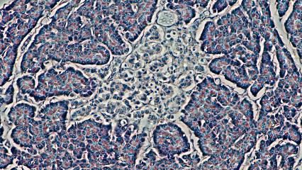
Replacing the insulin-producing cells lost in type 1 diabetes is only part of the challenge. The new cells must also survive in a body whose immune system may attack them. A research project beginning this month asks whether an injectable gel can help change that relationship. Breakthrough T1D lists a three-year award to Drexel University researcher Christopher Rodell, running from 1 November 2026 to 31 October 2029. The project concerns ToleroGel, a material designed to carry transplanted beta cells and a local immune-calming signal. This is a newly funded research programme, rather than a report of a cure in people. Two immune problems, one transplant In type 1 diabetes, immune activity destroys beta cells, which normally produce insulin in response to glucose. A transplant of donor-derived cells creates an additional issue: the recipient can recognise those cells as foreign. Protecting a graft therefore means addressing both the original autoimmune response and rejection of donor tissue. Rodell's proposal describes attaching the checkpoint protein B7x to a gel. The aim is to encourage protective regulatory immune cells around the transplant. Planned experiments include mouse models representing autoimmune diabetes and genetically mismatched donor cells. Success in either model would still require careful interpretation before translation to humans. Why protection is a consequential question NIDDK's patient guidance explains that donor-islet transplantation can improve blood-glucose control and reduce severe low-blood-sugar episodes in selected people. Islets contain several cell types, including beta cells. The procedure aims to restore biological insulin production rather than simply deliver insulin from outside the body. But recipients generally need medicines that prevent rejection for as long as their transplanted donor islets function. These medicines can increase susceptibility to infection and carry other serious risks. The balance is especially important when considering an intervention for a person already managing a lifelong condition. A local protection strategy would be valuable if it reduced that burden while preserving a functioning graft. That conditional statement is the reason for testing the idea. It should not be mistaken for evidence that the new material already provides safe, lasting protection. Existing cell therapies provide context In June 2023, the FDA approved Lantidra, made from deceased-donor pancreatic islets, for a limited group of adults with recurrent severe hypoglycaemia despite intensive management and education. The approval did not make islet transplantation a universal treatment for type 1 diabetes. The FDA's assessment described both insulin independence in some participants and serious adverse reactions associated with the infusion procedure and immunosuppression. That history shows why replacing cells and protecting them cannot be evaluated separately. A working graft is important, but the whole treatment pathway must be tolerable. Stem-cell-derived islets offer another route to obtaining replacement cells. A 2025 trial report found insulin independence at one year in ten of twelve full-dose participants. Those interim findings came from a small study using immunosuppression. They are separate evidence, not results for ToleroGel. How to read the project's promises The funder's project page sets out anticipated outcomes, including durable protection after the gel has degraded. Readers should recognise the grammatical difference between an aim and a finding. A three-year plan describes what investigators intend to test, even when its rationale is persuasive. Graymedy's interpretation is that durability will be particularly informative. If a material works only while a signal remains locally present, that raises a different development problem from establishing a lasting change in immune behaviour. Either result could teach researchers something useful, but they would support different claims. Other questions include whether the cells remain responsive to glucose, whether protection is reproducible and whether safety can be maintained while immune responses change. These are evaluation questions suggested by the goal of transplantation, rather than outcomes demonstrated by this newly started award. What would count as the next advance? A convincing next report would state the model used, the comparison treatment, how long graft function persisted and what harms were observed. Subsequent work would need to establish a reliable product and an appropriate route into human studies. A positive animal result would be a step in that process. For families, the useful update is that a named researcher is tackling a specific obstacle to cell replacement. Current insulin and diabetes-care plans remain matters for the treating team. Hope here rests on a testable question: can new cells be accepted without requiring the same level of systemic immune suppression? Sources: Breakthrough T1D: Rodell project and aims; NIDDK: islet transplantation; FDA: Lantidra approval; Stem-cell-derived islet trial, 2025. Photo: Berkshire Community College Bioscience Image Library / Wikimedia Commons, CC0. Reviewed 5 November 2026. Independently written from public sources; no interview was conducted. General information, not individual medical advice.
The coronavirus RNA map revealing new places to interrupt infection
Transplant research looks at the drainage system around the immune response